Solutions For Chronic Foot Pain
Painful conditions are considered chronic when symptoms have not resolved within twelve weeks despite treatment and medication. Foot and ankle pain usually persist when there is an underlying condition that has not been diagnosed.
Chronic Foot Pain May Be Multifocal
Single etiology pain is localized at the deformity (bunion, hammertoe, heel spur, neuroma, arthritis, etc). When the etiology of the deformity is straightforward, initial treatment should be effective. Pain is resolved and there is no ongoing problem.
When a patient presents with an obvious physical deformity, such as a bunion, hammertoe or a heel spur on x-ray, but they can't pinpoint where it hurts, this is a clue that there may be another issue that needs to be investigated.
Secondary conditions can drive vague, generalized or radiating complaints of foot and ankle pain. In some cases, pain isn't even generated from problems in the foot or ankle. If the patient has a history of low back pain or has developed degenerative disc disease, nerve roots can become compressed in the spinal column. Pain isn't only felt in the lower back. A painful electrical, shooting, and tingling sensation radiates from the tip of the toes to the back of their thighs or buttocks. Due to the location of the pain, patients usually assume they have a medical condition in the toe or foot.
If someone has undiagnosed diabetes, they may have peripheral neuropathy. Peripheral neuropathy presents with electrical tingling and burning sensations in the feet and is usually bilateral and symmetric. Burning sensation can also occur from poor blood circulation in the foot and ankle as a result of peripheral arterial disease (PAD).
How Can You Avoid Chronic Foot Pain?
Musculoskeletal problems of the foot and ankle are the primary reason patients seek initial treatment and are a large contributor to chronic pain. But if pain is generalized with the specific structural deformity, your doctor should be diligent in their initial examination. Unless all of the multifaceted components of your condition are identified and every component addressed, pain will persist after the initial treatment and chronic foot pain won't resolve.
Below are several in-office diagnostic tests your doctor can choose from to diagnose multifactorial problems when symptoms indicate.
Digital X-Ray
Digital x-ray is much clearer and more accurate than in the past when x-ray film was used. It is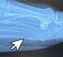 available in-office and is quick, painless and can be read in real time by your foot specialist. Viewing an x-ray image using a computer screen allows your doctor to enhance the image in a variety of ways to locate even small stress fractures. Radiographic imaging is used to diagnose chronic foot pain when the patient is hurting around the joints or in the bone.
Chronic pain conditions that can be diagnosed with in-office digital x-ray include:
Osteoarthritis
Rheumatoid Arthritis
Bone Spurs
Bone Fractures
Bone Deformity
Dislocated Joints
Infections
Osteoporosis
Bone Tumors
Bone Cysts
Musculoskeletal Ultrasound
Ultrasound is another in-office imaging procedure and uses sound waves to produce pictures of muscles, tendons, ligaments, nerves, and joints of the foot and ankle. It is commonly used to diagnosis pain as a resulting from sprains, strains, tears, trapped nerves, ganglion cysts and arthritis. It is painless, noninvasive and captures images in real time. Ultrasound is used instead of or in addition to digital x-ray when soft-tissue injury is suspected.
Chronic pain conditions that can be diagnosed with ultrasound include,
Plantar Fasciitis
Tenosynovitis
Tendon tear
Tendon rupture
Ligament tear
Ligament rupture
Capsulitis
Neuroma (Morton's Neuroma)
Ganglion Cyst
Entrapped Nerve
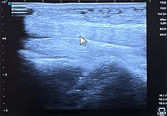
Healthy Tendon
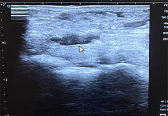
Torn Tendon
Arterial Doppler Study
An Arterial Doppler Study is done when signs point to poor blood circulation in the foot and ankle, such as pain in the foot, ankle and leg which worsens upon activity. Doppler is a third type of in-office imaging study that also uses sound waves in real time, but this diagnostic test uses ultrasound to listen to the blood flowing through the arteries and veins of your foot and ankle. The exam is painless, there are no needles, and the test has no side effects. Recordings of the arterial flow in the foot and ankle will be taken by placing a small probe over the dorsalis pedis artery on top of your foot and over the posterior tibial artery at the back of your ankle.
This study will detect if the arterial blood flow to the foot is not normal or it is diminished.
Blood Tests
 Most common lab tests ordered by a Foot and Ankle Specialist to diagnose chronic foot pain include:
- Complete Blood Count (CBC) with differential: tests for infection, osteomyelitis (bone infection)
- Erythrocyte Sedimentation Rate (ESR) : tests for inflammation in your body
- Uric Acid: tests for Gout (a type of inflammatory arthritis)
- Hemoglobin A1C: tells your doctor what your average blood glucose has been for the last 60 days. An elevated Hemoglobin A1C indicated uncontrolled blood sugar which can lead to nerve damage (neuropathy) causing chronic pain.
- Arthritis Panel: checks for autoimmune arthropathies causing joint cartilage damage.
Some of these tests are ordered together to assist with ruling out some conditions as well as confirming others. If, for example, you have pain in your big toe joint. Its red, hot, swollen and excruciatingly painful. You have not had an injury. Digital x-rays are clear. No ultrasound was done because the pain is in your toe joint. Arterial Doppler was not done because your dorsalis pedis and posterior tibial artery pulses are normal. Your doctor orders the following blood tests; CBC with Diff, ESR and Uric Acid. The tests come back within two days. The ESR is normal so there is no inflammation, the CBC is normal so there is no bone infection, the Uric Acid is elevated (greater than 8). So we know that you have had an acute gouty attack. Now your doctor can treat the cause and provide relief.
Biomechanical Analysis Of Foot And Ankle Function
The foot is a complex structure of 26 different bones, 33 joints, more than 100 muscles, tendons and ligaments, 2 main arteries and 2 main veins with hundreds of smaller veins providing blood flow and over 7,000 nerve endings - in each foot.
One quarter of all the bones in the human body are in the feet. The average person takes 8,000 to 10,000 steps a day with the push energy of walking adding another 30% of force to the person's standing body weight on the foot and ankle structures.
Biomechanical Analysis is advised when foot or ankle pain has lasted more than 30 days.
Your physician completes a thorough examination of how your joints, muscles, ligaments and tendons move together as you stand, walk and relax. It includes an ankle, forefoot and toe assessment, both non-weight bearing, then standing and walking. Range of motion, muscle strength and weakness, stability and flexibility, foot posture and a general posture assessment are completed.
The results provide your doctor with a complete understanding of how your foot and ankle functions, how your injuries and pain are occurring and what should be done to put your foot and ankle back into a healthy alignment to resolve pressure forces putting a strain on the structures of your foot and ankle.
Pathologies such as heel spurs, hallux valgus, hallux limitus, neuromas, shin splints, non-specific knee pain and non-healing diabetic wounds result from abnormal mechanics of the foot and ankle. Structural abnormalities involving the forefoot, midfoot and hindfoot impact center of foot pressure. Deviating gait motion with foot switch contact patterns, ankle/subtalar joint motion and center of foot pressure distribution strain joints, tendons, ligaments, muscles and nerves with each step.
Conservative and Surgical Treatment Options For Common Chronic Foot Pain Conditions
You can learn more about treatment options for specific conditions by visiting our Foot & Ankle Pain pages below.
- Persistent pain from Tendonitis; chronic inflammation of the foot or ankle tendons such as peroneal tendonitis
Make an Appointment